S. Burt Wolbach Photographs, 1918.
Dublin Core
Title
S. Burt Wolbach Photographs, 1918.
Subject
Spanish Influenza Epidemic, 1918-1919
Camp Devens (Mass.)
Wolbach, S. Burt (Simeon Burt), 1880-1954
Description
Wolbach, S. Burt (Simeon Burt), 1800-1954, M.D. Harvard Medical School, Boston, Massachusetts, 1903; was Shattuck Professor of Pathological Anatomy at Harvard Medical School from 1922 to 1947; pathologist-in-chief at Peter Bent Brigham, Boston Lying-in, and Children's Hospitals in Boston, Mass., until 1947; and director of Division of Nutritional Research at Children's Hospital from 1947 until 1954. His research was on infectious diseases, vitamin deficiency, and experimental pathology.
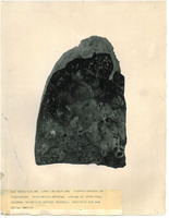
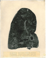
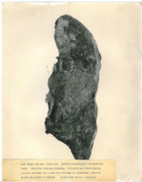
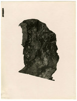
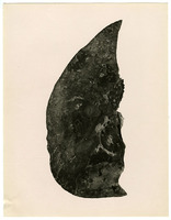
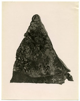
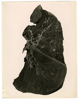
This collection features images of the lung slides created by Dr. S. Burt Wolbach at Camp Devens military base during the 1918 Influenza outbreak. When Wolbach arrived at Camp Devens there were 45,000 people and the base hospital was overwhelmed with influenza patients. Wolbach recorded twenty-eight postmortems at Camp Devens. He observed that there were two different stages of the disease: the lungs of patients who had died only a few days after the initial onset of symptoms had a different pathological profile than those who had lived ten days or longer with the illness. Wolbach published his initial findings in the April 1919 Johns Hopkins Bulletin and a more complete analysis of his research in the Archives of Internal Medicine in 1923.
This collection features images of the lung slides created by Dr. S. Burt Wolbach at Camp Devens military base during the 1918 Influenza outbreak. When Wolbach arrived at Camp Devens there were 45,000 people and the base hospital was overwhelmed with influenza patients. Wolbach recorded twenty-eight postmortems at Camp Devens. He observed that there were two different stages of the disease: the lungs of patients who had died only a few days after the initial onset of symptoms had a different pathological profile than those who had lived ten days or longer with the illness. Wolbach published his initial findings in the April 1919 Johns Hopkins Bulletin and a more complete analysis of his research in the Archives of Internal Medicine in 1923.
Is Part Of
Warren Anatomical Museum Records, Series 00344: V. Curatorial Photographs and Assorted Graphic Materials, 1973-1981, undated
Extent
20 photographs
Medium
Photographs
Language
English
Collection Items
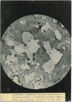
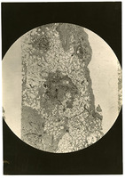
Photograph of a microscope slide from Camp Devens Case 192
An image of a microscope slide showing acute alveolar emphysema with hyalin fibrin deposit, which is characteristic of the initial stage of influenza pneumonias. The duration of the illness was 10 days from the initial symptoms.
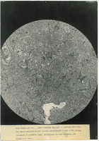
Photograph of a microscope slide from Camp Devens Case 214
An image of a microscope slide with the caption "CAMP DEVENS CASE 214. LOBAR PNEUMONIA SECONDARY TO INFLUENZA PNEUMONIA. / THE HYALIN FIBRINOUS DEPOSIT OUTLYING EMPHYSEMATOUS ALVEOLI OF THE INITIAL / INVOLVEMENT IS DISTINCTLY SEEN. BACTERIOLOGY…
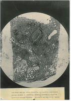
Photograph of a microscope slide from Camp Devens Case 219
An image of a microscope slide with the caption, "CAMP DEVENS CASE 219. CHRONIC BRONCHITIS, WITH ORGANIZING PERIBRONCHITIS. / ALVEOLAR EMPHYSEMA AND INTERSTITIAL EMPHYSEMA. EMPHYSEMA OF MEDIASTINUM / BACTERIOLOGY CABILLUS INFLUENZE. DURATION 21 DAYS…
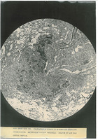
Photograph of a microscope slide from Camp Devens Case 223
An image of a microscope slide with the caption, "CAMP DEVENS CASE 223. ORGANIZATION OF EXUDATE IN BRONCHUS AND ORGANIZING / PERIBRONCHITIS. BACTERIOLOGY BACILLUS INFLUENZAE. DURATION 22 DAYS FROM / INITIAL SYMPTOMS."
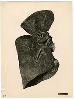
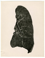
A photograph of the right lung from Camp Devens Case 223
A photograph of the lower lobe of a right lung with the caption, "CAMP DEVENS CASE 223. LOWER LOBE RIGHT LUNG. EXTENSIVE NECROSIS AND / ORGANIZATIONS. BRONCHIECTATIC ABSCESSES. ALVEOLAR AND INTERSTITIAL / EMPHYSEMA. BACTERIOLOGY BACILLUS INFLUENZAE.…
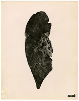
Photograph of a lung slide from Camp Devens Case 223
An image of a lung slide with the caption, "CAMP DEVENS CASE 223. CHRONIC BRONCHITIS. BRONCHIECTASES, PERIBRONCHITIS / AND BRONCHOPNEUMONIA. THE ORIGINAL ALVEOLAR EMPHYSEMA IS SHOWN AT APEX. / BACTERIOLOGY BACILLUS INFLUENZAE. DURATION 23 DAYS FROM…
Photograph of a right lung from Camp Devens Case 224
An image of a right lung slide with the caption, "CAMP DEVENS CASE 224. RIGHT LUNG. EXTENSIVE ORGANIZATION WITH BRONCHIEC- / TASES. ORGANIZING BRONCHIAL PNEUMONIA. BRONCHITIS AND PERIBRONCHITIS. / ALVEOLAR EMPHYSEMA FROM A CASE WITH EMPHYSEMA OF…
Photograph of a microscope slide from Camp Devens Case 193
An image of a microscope slide from a patient who died of influenza
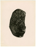
Photograph of a lung slide from Camp Devens Case 218
An image of a slide of a full lung that shows organizing bronchopneumonia.
Photograph of a rectus muscle slide from Camp Devens Case 216
An image of a rectus muscle slide. The muscle was ruptured and hemorrhaged.
Photograph of a lower lung from Camp Devens Case 198
An image of the lower lobe of a lung showing confluent bronchopneumonia
Photograph of a microscope slide from Camp Devens Case 183
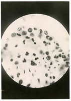
An image of a microscope slide showing clumps of influenza bacillia in the mucus membrane of a lung
Photograph of a right lung from Camp Devens Case 195
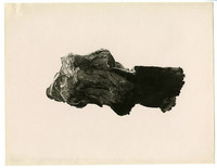
An image of a portion of a right lung with secondary pneumonia
Photograph of the lower lobe of a right lung from Camp Devens Case 212
An image of the lower lobe of a right lung with gangrene following the formation of a bronchiectatic abcess
Photograph of a lung from Camp Devens Case 225
An image of a lung showing lobar pneumonia following influenza pneumonia
Photograph of a lung slide from Camp Devens Case 198
An image of a lung slide showing signs of chronic bronchitis and peribronchitis not affected by the initial lesions
Photograph of the upper lobe of a right lung from Camp Devens Case 211
An image of the upper lobe of a right lung showing chronic bronchitis, peribronchitis, alveolar emphysema, and thickening of the interlobular septa
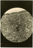
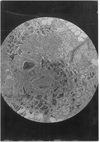
Photograph of a microscope slide from an unknown Camp Devens Case
An image of a microscope slide. This image is included with Wolbach's photographs from his research at Camp Devens, but no case number is recorded
Collection Tree
- Manuscripts and Archives
- Warren Anatomical Museum. Records, 1835-2010 (inclusive), 1971-1991. RG M-CL02.01. AA 192.5.
- S. Burt Wolbach Photographs, 1918.
- Warren Anatomical Museum. Records, 1835-2010 (inclusive), 1971-1991. RG M-CL02.01. AA 192.5.

