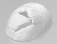
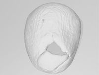
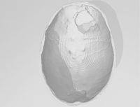
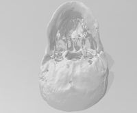
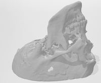
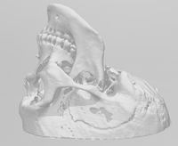
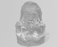
Phineas Gage (1823-1860) 3D STL Software Image
Dublin Core
Title
Phineas Gage (1823-1860) 3D STL Software Image
Subject
Gage, Phineas
Three-dimensional imaging
Description
Graham Holt (Director of the Office of Creative Solutions, Laboratories of Cognitive Neuroscience, Boston Children's Hospital) developed the initial Phineas Gage 3D print from CT scans taken by Peter Raitu and Ian Talos (Surgical Planning Laboratory, Brigham & Women’s Hospital). The 3D animation was printed with PLA plastic for the Beyond the Bone Box Collection, a Warren Anatomical Museum curated educational kit sponsored by Harvard Library S. T. Lee Innovation Grant. Gage's skull was separated into two separate files: top and bottom. The damage of the tamping iron accident caused bone damage, fragmentation, and missing pieces.
Abstract
The 3D image rendering of the Phineas Gage skull.
Creator
Graham Holt (Director of the Office of Creative Solutions, Laboratories of Cognitive Neuroscience, Boston Children's Hospital)
Format
model
Extent
1 photograph (3D animation)
Type
still image
Identifier
BoneBox_Gage_005_ref
Files
Citation
Graham Holt (Director of the Office of Creative Solutions, Laboratories of Cognitive Neuroscience, Boston Children's Hospital), “Phineas Gage (1823-1860) 3D STL Software Image,” OnView, accessed April 18, 2024, https://collections.countway.harvard.edu/onview/index.php/items/show/26404.

