Warren's Handbook
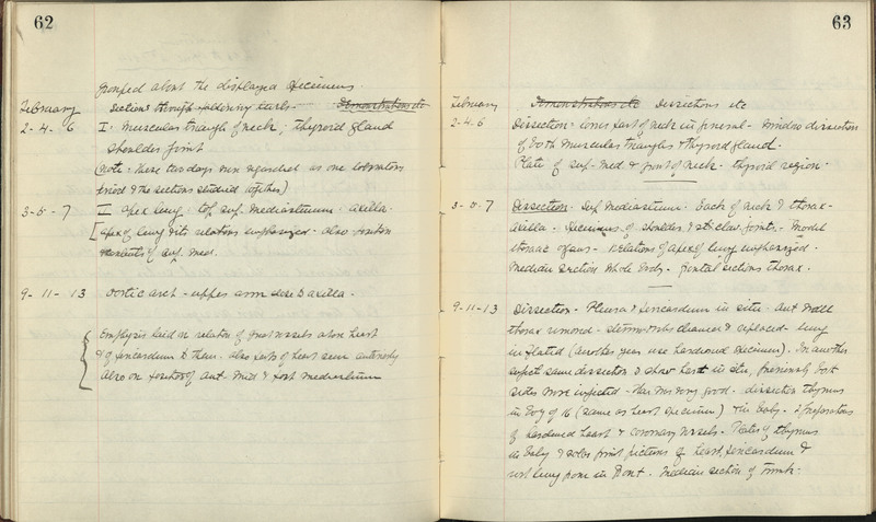
After Thomas Dwight’s death in 1911, the pre-eminent dissector at Harvard was John Warren (1874-1928), the great-great-grandson of the founder of the Medical School. This notebook was used by John Warren as Assistant, later Associate, Professor of Anatomy, to record the daily outline of lectures and dissections for first and second-year students, from 1911 to 1916. The pages displayed record Warren's notes on dissection exercises from February, 1914.
The lineal descendant of John Warren and John Collins Warren, John Warren (1874-1928) received a degree from the Medical School in 1900; he became a professor of anatomy at Harvard and was noted for his dissection work. During the last years of his life, Warren made over 400 preparations as teaching illustrations and commissioned artist Hamlet Frederick Aitken to reproduce them for a projected topographical atlas and handbook for dissection.
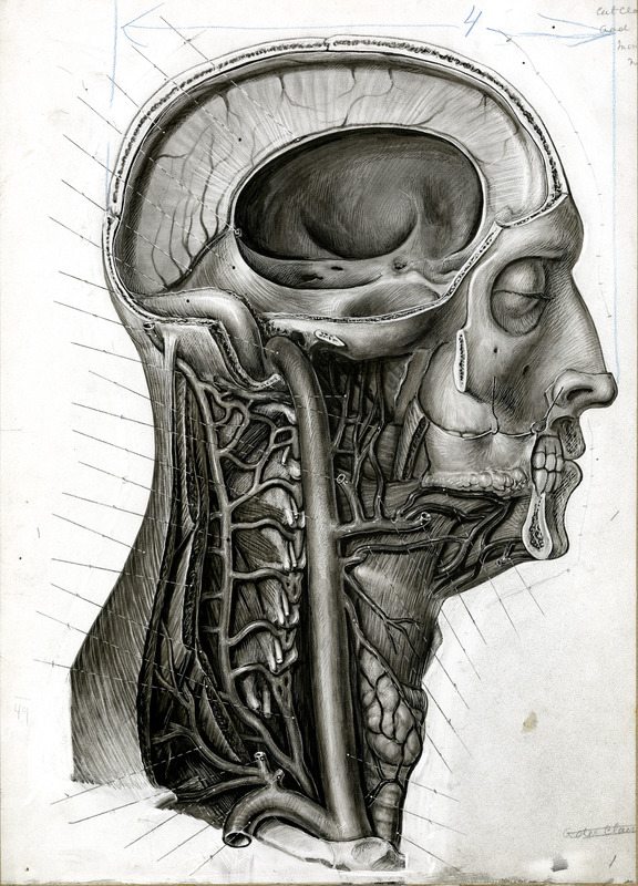
This is one of the several hundred original drawings produced by H. F. Aitken and eventually published in Warren's handbook. The text accompanying this illustration states, "The side of the skull has been cut away; the brain has been removed leaving the falx cerebri; the tunica mucosa oris is held up by hooks showing the glandula sublingualis; most of the clavicula is removed. The first rib, the trachea, glandula thyreoidea, and the cervical transverse processes, together with cut ends of the cervical nerves, are shown."
At Warren’s abrupt death, following complications from a fall, the preparations and illustrations were bequeathed to the Department of Anatomy. Robert Montraville Green (1880-1955), who taught gross and applied anatomy for nearly forty years, provided descriptive text and saw the project through to publication. Green edited Warren’s voluminous collection of drawings of dissections into publishable form and used that textbook to supplement his own lectures and demonstrations. Of Green’s work his memorial minute says, “The significant contact with the class was the lecture preceding each day’s dissection. The prosected and carefully draped cadaver was wheeled in, suggesting most strongly a patient in an old-fashioned surgical amphitheatre. Dr. Green … would demonstrate each part to be seen in the day’s work…. His attitude suggested that man was indeed a miracle, and that the students and professors were privileged as it were, to peer over the shoulder of God to view His handiwork in the structure of the human body.”
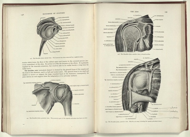
This copy of Warren's handbook belonged to Green and contains some of his emendations.


