Henry Jacob Bigelow Watercolor Collection, 1848-1854. WAM 21142.075—
Dublin Core
Title
Henry Jacob Bigelow Watercolor Collection, 1848-1854. WAM 21142.075—
Subject
Bigelow, Henry Jacob, 1818-1890
Wallis, Oscar
Teaching—Aids and devices
Description
Henry Jacob Bigelow was born 11 March 1818, in Boston, Massachusetts. His father, Jacob Bigelow (1787-1879), was a well-known medical practitioner in the city. Bigelow attended Harvard College, entering at the age of fifteen in 1833, and graduated with a B.A. in 1837. Afterwards, he began studying medicine with his father and attended lectures at Dartmouth College, Hanover, New Hampshire, given by Oliver Wendell Holmes (1809-1894). Bigelow was appointed a house surgeon at the Massachusetts General Hospital, Boston in 1838 and held the post for a year. He traveled to Cuba in the early 1840s and returned to Boston to take his medical degree in 1841. After completing his M.D., Bigelow traveled to Europe, studying medicine in Paris and London until his return to Boston in 1844 to take up medical practice. In the same year, he wrote the Boylston Prize-winning essay on that year’s topic of the relief of deformity and lameness by muscle division. In 1845, he was elected president of the Boylston Society and in 1846, appointed Visiting Surgeon in the Massachusetts General Hospital. In 1849, Bigelow was appointed as Professor of Surgery at the Harvard Medical School. Bigelow was a pathologist by training, but was active in a variety of other fields, including the popularizing of anesthesia by ether, revising the equipment and technique for lithotrity, and working with the anti-vivisection movement. He was a supporter of the claim of William Morton as the discoverer of anesthetic ether and wrote widely on the subject; Bigelow also favored the immediate adoption of ether anesthetic in surgical operations, claiming to have been responsible for one of the first uses of the discovery in surgery at the Massachusetts General Hospital. Bigelow worked with artist Oscar Wallis on a detailed series of medical illustrations of tumors and other specimens viewed under the
microscope. Other drawings on which Bigelow and Wallis collaborated were used in Bigelow's published works or as teaching tools, but the drawings in this collection were mostly intended for a work which was never published. Henry Jacob Bigelow died in 1890 at his home in Newton, Massachusetts, several months after a severe fall.
Oscar Wallis was an artist primarily known for his 1848-1854 collaboration with Henry Jacob Bigelow for a set of teaching watercolors. Wallis also illustrated a volume on pathology written by Bigelow, although this volume was never published. Wallis also contributed lithographic work to Benjamin Champney's "New England Scenery" (1852) and George Bond's "Map of the White Mountains" (1853).
This collection is comprised of the set of watercolor illustrations that Henry Jacob Bigelow employed artist Oscar Wallis to paint from 1848-1854. Bigelow commissioned these large watercolors of anatomical subjects to illustrate his lectures at Harvard Medical School. Wallis painted the teaching diagrams from local subjects and from the atlases of established medical authorities.
Provenance
In 1890 Bigelow presented the watercolors to Reginald H. Fitz to be used in the Harvard Medical School's Department of Anatomy. The watercolors were transferred into the Warren Anatomical Museum between 1890 and 1930.
Collection Items
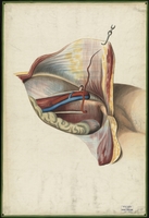
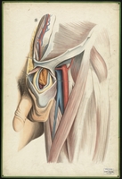
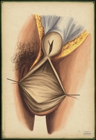
Teaching watercolor of the structure of the crural ring in the groin
After Thomas George Morton's The surgical anatomy of the principal regions of the human body, The Groin, plate 5, figure 1
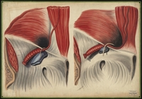
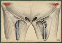
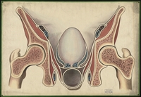
Teaching watercolor showing the circular dilation of the internal abdominal ring and the changed position of the epigastric artery caused by an inguinal hernia
After Thomas George Morton's The surgical anatomy of the principal regions of the human body, Inguinal Herniae, figures no. 4 and 5
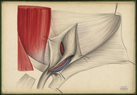
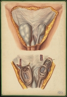
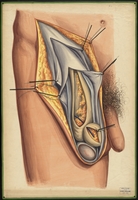
Teaching watercolor of the sheath of the fascia transversalis of the spermatic cord
After Thomas George Morton's The surgical anatomy of the principal regions of the human body, Inguinal Herniae, figure no. 2
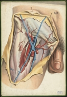
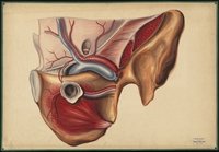
Teaching watercolor of major vessels of the hip
After Thomas George Morton's The surgical anatomy of the principal regions of the human body, The Groin, nos. 3 and 6
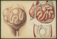
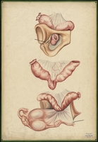
Teaching watercolor of umbilical hernias
After Jean Cruveilhier's Anatomie pathologique du corps humain, vol. 2, liv. 24, pl. 6
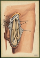
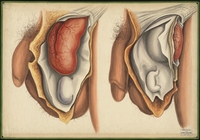
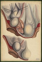
Teaching watercolor of inguinal hernias in male patients
After Jean Baptiste Marc Bourgery's Traite complet de l'anatomie de l'homme, plate 34
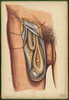
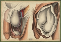
Teaching watercolor of indirect and direct inguinal hernias
After Jean Baptiste Marc Bourgery's Traite complet de l'anatomie de l'homme, plate 38 bis.
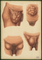
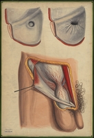
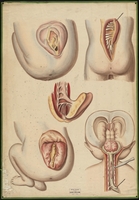
Teaching watercolor of the external signs of various types of hernias in females
After Jean Baptiste Marc Bourgery's Traite complet de l'anatomie de l'homme, vol. 7, plate 36
Teaching watercolor of an existing congenital hernia on the same side and in the same sheath as an accidental hernia
After J. B. D. Demeaux's Recherches sur l'evolution du sac herniaire suivies de considerations chirurgicales sur les complications auxelles il pent donner lieu, plate 7
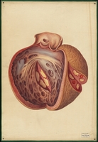
Teaching watercolor of hernia in the testis
After J. B. D. Demeaux's Recherches sur l'evolution du sac herniaire suivies de considerations chirurgicales sur les complications auxelles il pent donner lieu, plate 8
Teaching watercolor showing an oblique inguinal hernia where the two sacs are beside each other
After J. B. D. Demeaux's Recherches sur l'evolution du sac herniaire suivies de considerations chirurgicales sur les complications auxelles il pent donner lieu, plate 4
Teaching watercolor showing an inguinal hernia where one sac is within the wall of the other
After J. B. D. Demeaux's Recherches sur l'evolution du sac herniaire suivies de considerations chirurgicales sur les complications auxelles il pent donner lieu, plate 6
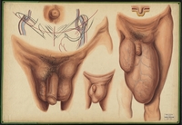
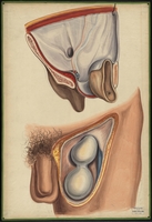
Teaching watercolor of a congenital hernia and an infantile hernia in male subjects
After Thomas George Morton's The surgical anatomy of the principal regions of the human body, Inguinal Herniae, pages 281 and 284
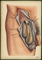
Teaching watercolor of the tunica vaginalis and the vessels of the spermatic cord from a scrotal hernia
After Thomas George Morton's The surgical anatomy of the principal regions of the human body, Inguinal Herniae, figures no. 6 and 11
Teaching watercolor of inguinal hernia
After J. B. D. Demeaux's Recherches sur l'evolution du sac herniaire suivies de considerations chirurgicales sur les complications auxelles il pent donner lieu, plate 1
Teaching watercolor of the coverings of the sac of an oblique or external inguinal hernia
After Thomas George Morton's The surgical anatomy of the principal regions of the human body, Inguinal Herniae, plate 5
Teaching watercolor of a dissected inguinal hernia sacs in a female
After J. B. D. Demeaux's Recherches sur l'evolution du sac herniaire suivies de considerations chirurgicales sur les complications auxelles il pent donner lieu, plate 3
Teaching watercolor of the saphenous opening of the fascia lata and the saphena vein, with and without the cribriform fascia
After Thomas George Morton's The surgical anatomy of the principal regions of the human body, The Groin, pages 102 and 104
Teaching watercolor of a femoral hernia in a female patient
After Thomas George Morton's The surgical anatomy of the principal regions of the human body, The Groin, plate 6
Teaching watercolor of inguinal hernia
After J. B. D. Demeaux's Recherches sur l'evolution du sac herniaire suivies de considerations chirurgicales sur les complications auxelles il pent donner lieu, plate 2
Teaching watercolor of a hernia of the uterus
After Jean Cruveilhier's Anatomie pathologique du corps humain, volume 2, part 34, plate 6
Teaching watercolor of obturator hernia
After Robert Druitt's The principles and practice of modern surgery, page 465
Teaching watercolor of two femoral hernias
After J. B. D. Demeaux's Recherches sur l'evolution du sac herniaire suivies de considerations chirurgicales sur les complications auxelles il pent donner lieu, plate 5
Teaching watercolor of femoral hernia of the intestines
After Jean Cruveilhier's Anatomie pathologique du corps humain, vol. 1, liv. 15, pl. 6
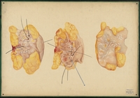
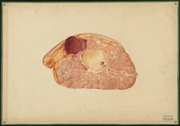
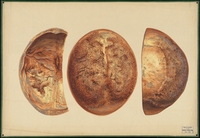
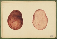
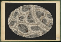
Teaching watercolor of cross sections of a piece of removed tissue
Possibly from a local Boston patient
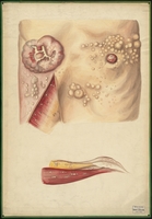
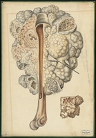
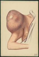
Large teaching watercolor of breast neoplasms in the skin and muscles of the breast
After Jean Cruveilhier's Anatomie pathologique du corps humain, vol. 2, liv. 26, plate 3
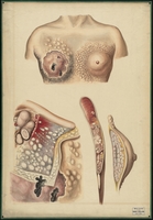
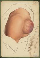
Teaching watercolor of advanced breast cancer
After Jean Cruveilhier's Anatomie pathologique du corps humain, vol. 2, liv. 31, pl. 2
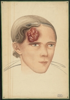
Teaching watercolor of a tumor on the forehead of a female patient
Possibly of a local Boston patient
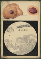
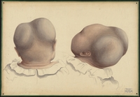
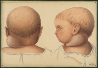
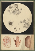
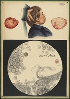
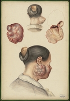
Teaching watercolor of a large growth on a child's head and a microscopic view of the tissue
Possibly of a local Boston patient
Teaching watercolor of the abnormal dilation of the subcutaneous abdominal veins
After Jean Cruveilhier's Anatomie pathologique du corps humain, vol. 1 liv. 16, plate 6
Teaching watercolor showing interior layers of coagular of an enormous aneurysm of the descending aorta, an aneurysm of the carotid, and the contraction of the aorta
After Jean Cruveilhier's Anatomie pathologique du corps humain, vol. 2, liv. 40, pl. 3
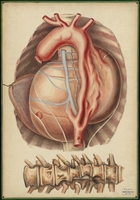
Teaching watercolor of enormous aneurysm of the descending aorta and spinal deterioration caused by contact with it
After Jean Cruveilhier's Anatomie pathologique du corps humain, vol. 2, liv. 40, pl. 2
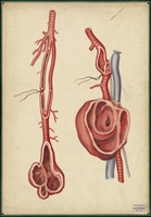
Teaching watercolor of an inguinal aneurysm and the femoral and popliteal arteries with a popliteal aneurysm
After Sir Charles Bell's Practical Essays, vol. 2, pl. 2, by Oscar Wallis
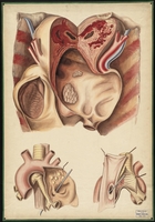
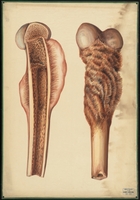
Teaching watercolor of an aneurysm of the aorta, trachea, and esophagus
After Jean Cruveilhier's Anatomie pathologique du corps humain, volume 1, part 3, plate 4
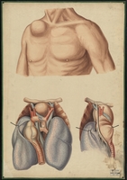
Teaching watercolor of an aneurysm of the aortic arch which pushed through the gap between the clavicle and the rib cage to protrude beneath the skin
After Jean Cruveilhier's Anatomie pathologique du corps humain, vol. 1, liv. 3, pl. 3
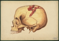
Teaching watercolor of an aneurysm by anastomosis on the skull
After Robert Druitt's The principles and practice of modern surgery, page 302
Teaching watercolor of five views of spinal dysraphism in infants
After Jean Cruveilhier's Anatomie pathologique du corps humain, volume 1, part 16, plate 4
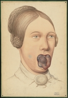
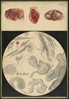
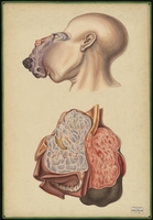
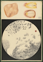
Teaching watercolor of a tumor in the mouth, including exterior views, dissected view, and view of tissue under a microscope
Possibly from a local Boston patient
Teaching watercolor of exterior and interior views of a large bone tumor of the upper jaw which filled the cavities of the nose and orbits and extended into the cranium
After Edward Stanley's Illustrations of the effects of disease and injury of the bones, plates 13 and 17
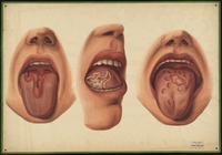
Teaching watercolor of tongue ulcers and papules caused by venereal disease
After Philippe Ricord's Traite complet des maladies veneriennes, plate 35
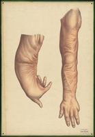
Teaching watercolor of a deformity of the arm caused by a severe burn, and the results of corrective surgery on the same arm
After Jacques D. Lebaudy's The anatomy of the regions interested in the surgical operations performed upon the human body, plate 18
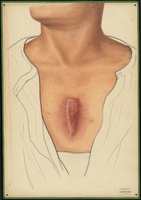
Teaching watercolor of an abnormal growth on the sternum of a male subject
Possibly of a local Boston patient
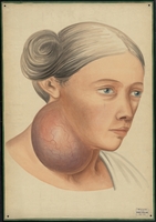
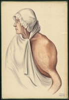
Teaching watercolor of woman with large growth on right side of neck under the ear and jaw
Possibly after a local Boston patient
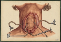
Teaching watercolor of the removal of a cancerous goiter
After Jean Baptiste Marc Bourgery's Traite complet de l'anatomie de l'homme, volume 7, plate 26, figure 4
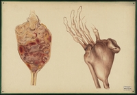
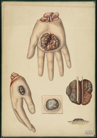
Teaching watercolor of a tumor removed from the ring finger of the left hand and a microscopic view of the tissue
Possibly from a local Boston patient
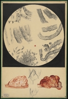
Teaching watercolor of removed skin tissue and a microscopic view of that tissue
Possibly from a local Boston patient
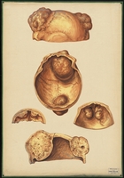
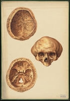
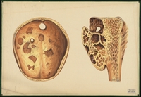
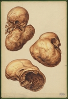
Teaching watercolor showing several views of tumors in the bone of the skull
Possibly after plate 15 of an unidentified source
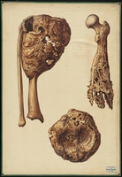
Teaching watercolor of a large tumor on the humerus
After Jean Cruveilhier's Anatomie pathologique du corps humain, vol. 2, liv. 34, pl. 4
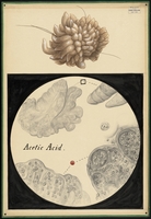
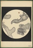
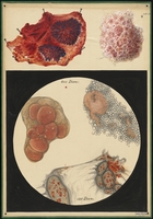
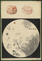
Teaching watercolor of a removed mass with a microscopic view of the tissue
Possibly from a local Boston patient
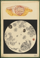
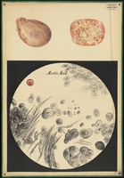
Teaching watercolor of a removed tumor and a microscopic view of the tissue
Possibly from a local Boston patient
Teaching watercolor of a woman with a large tumor on her humerus
After Edward Stanley's Illustrations of the effects of disease and injury of the bones, plate 13
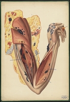
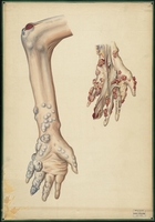
Teaching watercolor of tumors throughout the left arm
After Jean Cruveilhier's Anatomie pathologique du corps humain, vol. 2, liv. 30, pl. 5
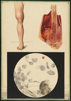
Teaching watercolor of a mass on the femur near the knee and a microscopic view of the tissue
Possibly from a local Boston patient
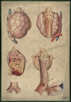
Teaching watercolor of diseases of the larynx, trachea, and thyroid gland
After Jean Cruveilhier's Anatomie pathologique du corps humain, vol. 2, liv. 35, pl. 4
Teaching watercolor of a mass removed from the jaw of a male child and a microscopic view of the tissue
Possibly of a local Boston patient
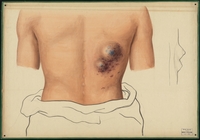
Teaching watercolor of large mass on the back of the left shoulder
Possibly of a local Boston patient
Teaching watercolor of tumors in the lower arm and hand
After Jean Cruveilhier's Anatomie pathologique du corps humain, vol. 2, liv. 23, pl. 3 and 4
Teaching watercolor of scar tissue, tumors, and abnormality of the nerves
After Jean Cruveilhier's Anatomie pathologique du corps humain, vol. 2, liv. 35, pl. 2
Teaching watercolor of removed masses in skin tissue and a microscopic view of the tissue
Possibly from local Boston patient
Teaching watercolor of the lower arm cut horizontally near the elbow to show muscles, bone, nerves, and blood vessels
After Alfred Velpeau's Traite complet d'anatomie chirurgicale, generale et topographique du corps humain, plate 17
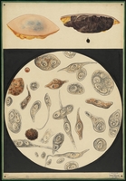
Teaching watercolor of removed tissue and a microscopic view of it
Possibly from a local Boston patient
Teaching watercolor of degeneration of the heart tissue caused by venereal disease
After Phillipe Ricord's Traite complet des maladies veneriennes, plate 29
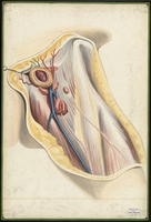
Teaching watercolor of the three-part sheath of the femoral vessels
After Thomas George Morton's The surgical anatomy of the principal regions of the human body, The Groin, plate 4
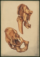
Teaching watercolor of bone loss of the cranium caused by bone cancer and of a tibia with late stage bone cancer
After Auguste Nelaton's Elemens de pathologie chirurgicale, fig. 243 and 244
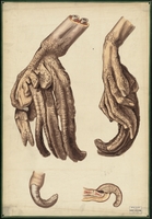
Teaching watercolor of a right hand with a skin disease that causes scaly skin and horn-like growths
After Jean Cruveilhier's Anatomie pathologique du corps humain, vol. 1, liv. 7, pl. 6
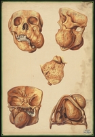
Teaching watercolor of five views of a tumor of the skull and jaw
Possibly of a local Boston patient
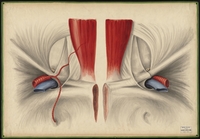
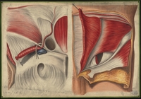
Teaching watercolor of the posterior wall of the inguinal canal and a view, from within, of the neck of the sac of a direct or internal inguinal hernia
After Thomas George Morton's The surgical anatomy of the principal regions of the human body, Inguinal Herniae, no. 3 and 9
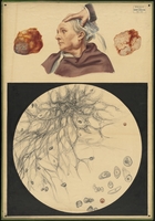
Teaching watercolor of a mass removed from the jaw of a woman with a microscopic view of the tissue
Possibly of a local Boston patient
Teaching watercolor of a cartilaginous tumor of the humerus
After Edward Stanley's Illustrations of the effects of disease and injury of the bones, plate 19
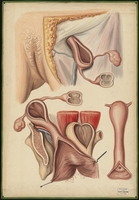
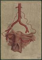
Teaching watercolor of the uterine, ovarian, and pudic arteries
After Richard Quain's The anatomy of the arteries of the human body and its applications to pathology and operative surgery, plate 63
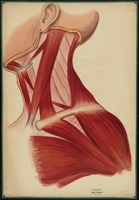
Teaching watercolor of the muscles of the neck, chest, jaw, and clavicle
AfterThomas Wormald's A series of anatomical sketches and diagrams, plate 6
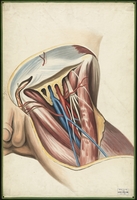
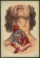
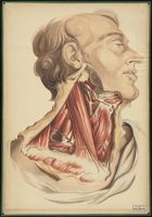
Teaching watercolor of a dissection of the neck showing muscles and blood vessels
Possibly after plate 17 of an unknown work
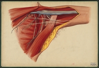
Teaching watercolor of the region of the Axilla on the left side following the removal of the adipose and cellular tissue
After Thomas Wormald's A series of anatomical sketches and diagrams, plate 17
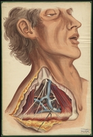
Teaching watercolor of the blood vessels in the right side of the neck
Possibly of local Boston patient
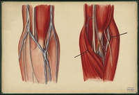
Teaching watercolor of the muscles, nerves, and blood vessels of the inner elbow
After Thomas Wormald's A series of anatomical sketches and diagrams, plate 18
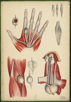
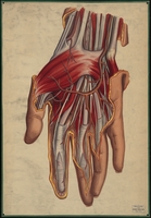
Teaching watercolor of a surgical dissection of the wrist and palm
After Joseph Maclise's Surgical Anatomy, III. 9
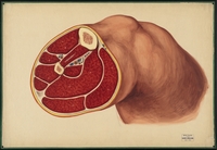
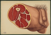
Teaching watercolor of the thigh cut horizontally at the midpoint to show muscles, bone, nerves, and blood vessels
After Alfred Velpeau's Traite complet d'anatomie chirurgicale, generale et topographique du corps humain, plate 16
Teaching watercolor of cancer on the hand
After Jean Cruveilhier's Anatomie pathologique du corps humain, vol. 1, liv. 19, pl. 3
Teaching watercolor of a bone tumor and a microscopic view of the tissue
Possibly from local Boston patient
Teaching watercolor of the appearance of vessels in the web of a frog's foot after the addition of water
After Addison's Physiological Researches, plate 2
Collection Tree
- Warren Anatomical Museum
- Henry Jacob Bigelow Watercolor Collection, 1848-1854. WAM 21142.075—

