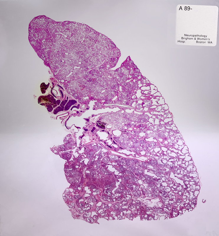
Lung Section
Human Lung Section, 1989
“This section, representing a lung lobe (15cm x 7cm), shows Usual Interstitial Pneumonia, the pathologic manifestation of Idiopathic Pulmonary Fibrosis. This is the most common form of idiopathic (unexplained) lung fibrosis in the adult population. This is an irreversible form of fibrosing lung disease that leads to progressive shortness of breath and is fatal. This is one lobe of an adult lung sampled at the time of autopsy. The tissue at the top of the lobe appears normal—the individual air sacs that make up the lung are thin-walled and barely visible. At the outer and lower edges of the lobe, however, the airspaces are visibly enlarged, surrounded by fibrotic tissue, and no longer support gas exchange. This change can be perceived on radiographic imaging as ‘honeycomb lung.’
In usual clinical practice, histology microtomes can create sections from tissue samples measuring at most 2 to 3 centimeters in size. This sample is remarkable for its size and architectural preservation. Creating this section required specialized equipment and histology technique.” —Lynette Marie Sholl, MD, Associate Pathologist, Brigham and Women’s Hospital, Associate Professor of Pathology, HMS

