
Browse Items (20 total)
- Collection: S. Burt Wolbach Photographs, 1918.
Sort by:
Photograph of the upper lobe of a right lung from Camp Devens Case 211
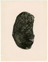
An image of the upper lobe of a right lung showing chronic bronchitis, peribronchitis, alveolar emphysema, and thickening of the interlobular septa
Photograph of the lower lobe of a right lung from Camp Devens Case 212
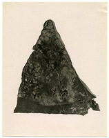
An image of the lower lobe of a right lung with gangrene following the formation of a bronchiectatic abcess
Photograph of the left lung from Camp Devens Case 211
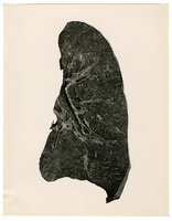
An image of a left lung
Photograph of a right lung from Camp Devens Case 224
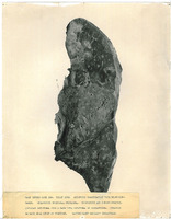
An image of a right lung slide with the caption, "CAMP DEVENS CASE 224. RIGHT LUNG. EXTENSIVE ORGANIZATION WITH BRONCHIEC- / TASES. ORGANIZING BRONCHIAL PNEUMONIA. BRONCHITIS AND PERIBRONCHITIS. / ALVEOLAR EMPHYSEMA FROM A CASE WITH EMPHYSEMA OF…
Photograph of a right lung from Camp Devens Case 195
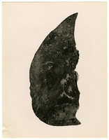
An image of a portion of a right lung with secondary pneumonia
Photograph of a rectus muscle slide from Camp Devens Case 216
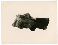
An image of a rectus muscle slide. The muscle was ruptured and hemorrhaged.
Photograph of a microscope slide from Camp Devens Case 223
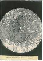
An image of a microscope slide with the caption, "CAMP DEVENS CASE 223. ORGANIZATION OF EXUDATE IN BRONCHUS AND ORGANIZING / PERIBRONCHITIS. BACTERIOLOGY BACILLUS INFLUENZAE. DURATION 22 DAYS FROM / INITIAL SYMPTOMS."
Photograph of a microscope slide from Camp Devens Case 219
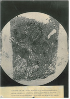
An image of a microscope slide with the caption, "CAMP DEVENS CASE 219. CHRONIC BRONCHITIS, WITH ORGANIZING PERIBRONCHITIS. / ALVEOLAR EMPHYSEMA AND INTERSTITIAL EMPHYSEMA. EMPHYSEMA OF MEDIASTINUM / BACTERIOLOGY CABILLUS INFLUENZE. DURATION 21 DAYS…
Photograph of a microscope slide from Camp Devens Case 214
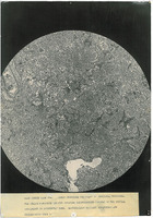
An image of a microscope slide with the caption "CAMP DEVENS CASE 214. LOBAR PNEUMONIA SECONDARY TO INFLUENZA PNEUMONIA. / THE HYALIN FIBRINOUS DEPOSIT OUTLYING EMPHYSEMATOUS ALVEOLI OF THE INITIAL / INVOLVEMENT IS DISTINCTLY SEEN. BACTERIOLOGY…
Photograph of a microscope slide from Camp Devens Case 193
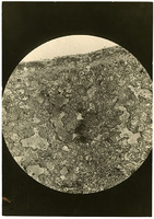
An image of a microscope slide from a patient who died of influenza
Photograph of a microscope slide from Camp Devens Case 192
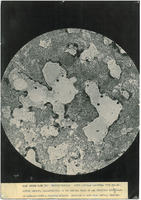
An image of a microscope slide showing acute alveolar emphysema with hyalin fibrin deposit, which is characteristic of the initial stage of influenza pneumonias. The duration of the illness was 10 days from the initial symptoms.
Photograph of a microscope slide from Camp Devens Case 183
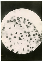
An image of a microscope slide showing clumps of influenza bacillia in the mucus membrane of a lung
Photograph of a microscope slide from an unknown Camp Devens Case
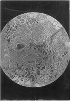
An image of a microscope slide. This image is included with Wolbach's photographs from his research at Camp Devens, but no case number is recorded
Photograph of a lung slide from Camp Devens Case 223
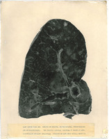
An image of a lung slide with the caption, "CAMP DEVENS CASE 223. CHRONIC BRONCHITIS. BRONCHIECTASES, PERIBRONCHITIS / AND BRONCHOPNEUMONIA. THE ORIGINAL ALVEOLAR EMPHYSEMA IS SHOWN AT APEX. / BACTERIOLOGY BACILLUS INFLUENZAE. DURATION 23 DAYS FROM…
Photograph of a lung slide from Camp Devens Case 218
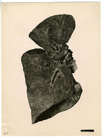
An image of a slide of a full lung that shows organizing bronchopneumonia.
Photograph of a lung slide from Camp Devens Case 204
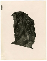
An image of a lung with interstitial emphysema
Photograph of a lung slide from Camp Devens Case 198
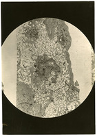
An image of a lung slide showing signs of chronic bronchitis and peribronchitis not affected by the initial lesions
Photograph of a lung from Camp Devens Case 225
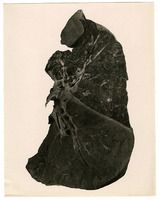
An image of a lung showing lobar pneumonia following influenza pneumonia
Photograph of a lower lung from Camp Devens Case 198
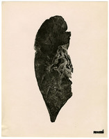
An image of the lower lobe of a lung showing confluent bronchopneumonia
A photograph of the right lung from Camp Devens Case 223
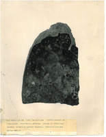
A photograph of the lower lobe of a right lung with the caption, "CAMP DEVENS CASE 223. LOWER LOBE RIGHT LUNG. EXTENSIVE NECROSIS AND / ORGANIZATIONS. BRONCHIECTATIC ABSCESSES. ALVEOLAR AND INTERSTITIAL / EMPHYSEMA. BACTERIOLOGY BACILLUS INFLUENZAE.…