Artifacts on Display
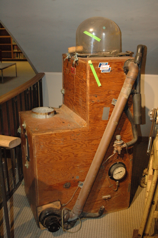
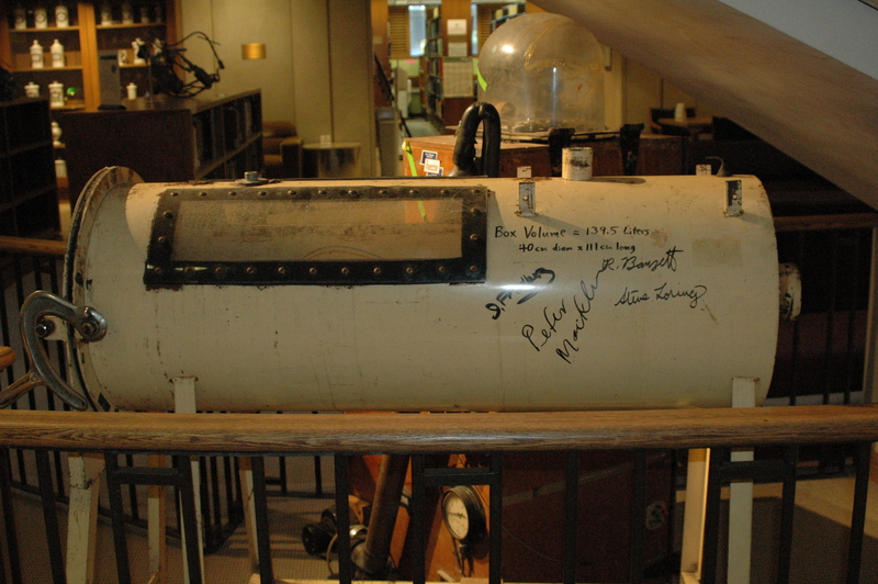
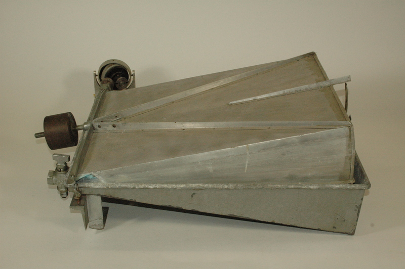
The two large devices, called plethysmographs, were used to measure breathing, lung gas volumes, and airway resistance in the Harvard School of Public Health’s Department of Physiology in the 1960s and 1970s. A human or animal subject was inside the plethysmographs’ sealed interior and breathed through a mouthpiece connected to the outside. At any lung volume, the mouthpiece could be closed. The subjects would then make inspiratory and expiratory efforts, causing the lungs to contract or expand. The smaller device is a spirometer. It would capture the corresponding changes in lung size by measuring the air leaving or entering the closed system. These devices were local School of Public Health adaptations in the larger field of mid-20th century scientific plethysmography. The data obtained expanded knowledge of lung physiology, growing the understanding of diseases with altered airway function and other pathologies caused by smoking and polluted air.
The volume displacement plethysmograph was first used to measure lung volumes, airway function, and rapid breathing events in human subjects by Harvard physiologist Jere Mead in 1960. Mead was building on Arthur DuBois and Julius Comroe, Jr.’s mid-1950s pioneering efforts on the plethysmographic method and thoracic gas volume and airway function measurement. The Mead body box was a smaller, more portable model than the initial DuBois-Comroe chamber, offering a more rapid response time with enhanced reproducibility.
This plethysmograph was used at the School of Public Health for research on the respiratory system between 1950 and 1990. The device resembles an iron lung and was designed to determine lung volumes and characterize breathing in anesthetized dogs.
A 1981 paper in the Journal of Applied Physiology entitled “Vagal Stimulation and Aerosol Histamine Increase Hysteresis of Lung Recoil” used this plethysmograph to explore the relationship between air flow resistance (an important feature of asthma) and the viscous behavior of lung tissue.
As a result of this study and other plethysmograph research, the Department of Environmental Health continues to deepen our understanding of lung physiology, which in turn shapes our understanding of air pollution and occupational dusts and their impact on human health.
In a 1968 study entitled “Pressure-Flow Events During Singing,” this plethysmograph recorded the breathing patterns of a singer. Measurements were made of lung volume changes, subglottic pressures, and airflow rates during the singing of tones of measured pitch and loudness. By broadening the understanding of respiratory mechanics during human activities, this research gave public health scientists better tools to measure how airborne environmental exposures might enter the body.


