Intubation: Laryngoscopes
Laryngoscopes
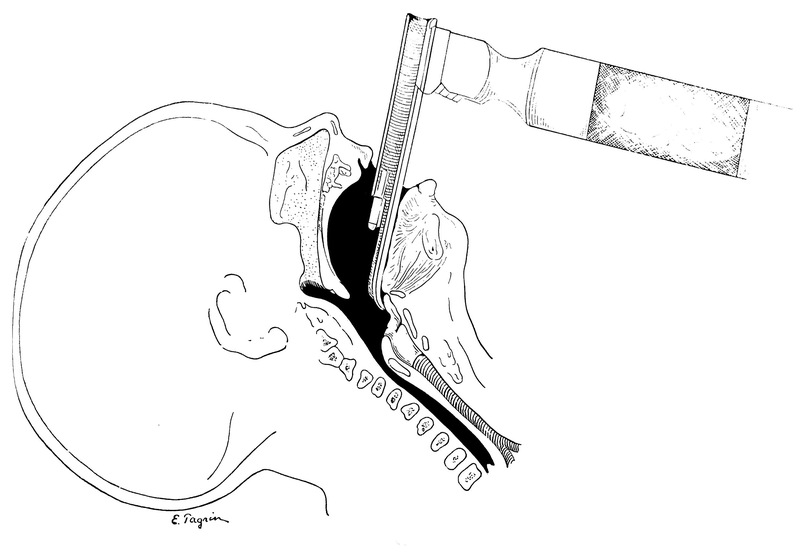
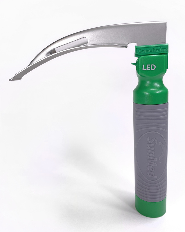
A laryngoscope consists of a handle, a blade, and a built-in light source. Anesthesiologists use them to help place endotracheal tubes in the trachea which allows them to control a patient’s airway for the administration of oxygen and anesthesia. In contemporary practice, the use of a mounted video camera connected to a display screen has become popular, often replacing traditional laryngoscopes in the operating room. The advantage of video is that it provides an excellent view of the vocal cords, especially with anatomically challenging airways. Some anesthesiologists choose video for every intubation.
From the 1850s through the 1930s many techniques for assisting with the placement of endotracheal tubes were explored (including just the doctor’s finger) until the ultimate refinement in the design of laryngoscope blades was introduced in the US in the early 1940s. The “Miller” with a long straight blade, was designed by Robert Miller in 1941. The “MacIntosh” with a short, curved blade, was designed by Robert MacIntosh in 1943 in the UK. All subsequent designs are small variations of these two.
“Between the Macintosh (curved and shorter) and Miller (straight and longer) blades, the Macintosh is more popular because it is less stimulating (it is not inserted as deeply into the airway) and offers good viewing conditions for intubation in most patients. However, in some patients it is advantageous to insert the laryngoscope deeper, lifting a structure called the epiglottis, to get a better view of the larynx. For such patients, and for individuals whose training institutions used Miller blades, this would be the first one chosen. Moreover, the Miller blade is often preferred in children. At the Brigham and probably in most US hospitals, the Macintosh blade is used preferentially.” —Sukumar Desai, MD, BWH Department of Anesthesiology, Perioperative and Pain Medicine
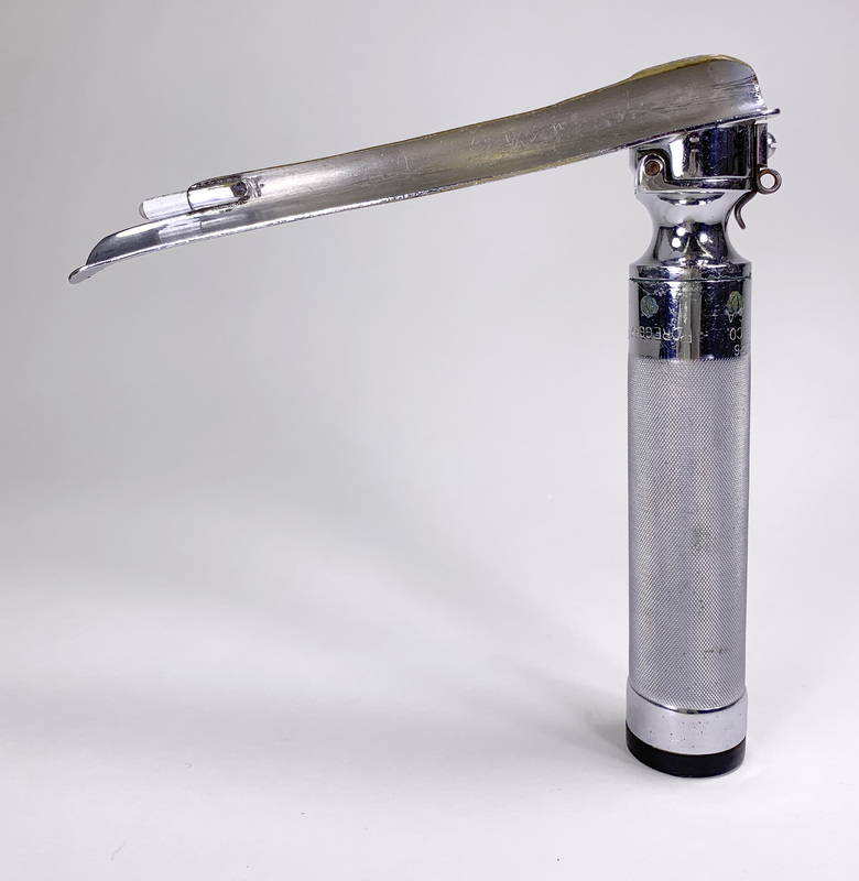
Foregger Laryngeal Speculum, Miller Style, circa 1940s
This example is an early improvement to the Miller designed straight-bladed laryngoscope. Richard Foregger’s version included a collapsible blade. His idea was granted a patent in 1942.
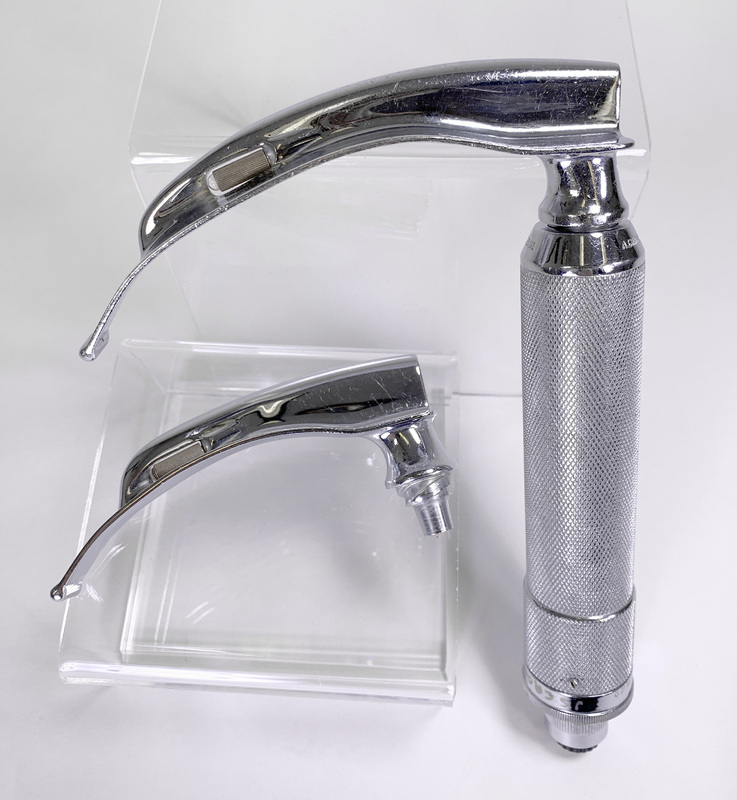
Macintosh Style Laryngoscope with Quick Screw Base, circa 1950s
(Style introduced 1943; Base patented 1950)
The blade of this laryngoscope is detachable from the body by a quick screw fitting into a threaded recess in the body allowing fast blade size changes. This example includes a 150mm blade (large adult) and a 120mm blade (adult medium).


