Electron Microscopes
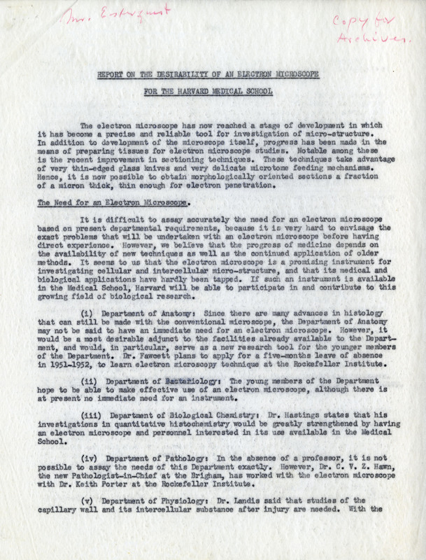
Under the chairmanship of George Bernays Wislocki (1892-1956), the Hersey Professor from 1947 to 1956, the focus of the Department of Anatomy began to shift to embryology and histology, and the funding for the first electron microscope at the Medical School was obtained in the 1953-54 academic year. The next Hersey Professor, Don Wayne Fawcett (1917-2009), encouraged research with the electron microscope, and the anatomical course description for the first-years reflects this. Students were to gain
…familiarity with the normal structure of cells and tissues as they appear under light microscope and with their finer structure as revealed by the electron microscope. The histology presented in the second semester forms part of the correlative teaching in which the structure, physiology, and biochemistry of the major organ systems are studied. Demonstrations of fresh tissue are prepared for examination with phase contrast and interference microscopy, and special cytological and histochemical preparations are displayed to present the broad range of techniques used in morphological investigations. Embryology is taught in conjunction with the principles of genetics in a series of lectures extending throughout the first semester.
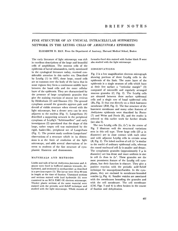
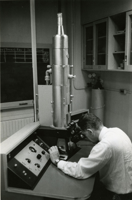
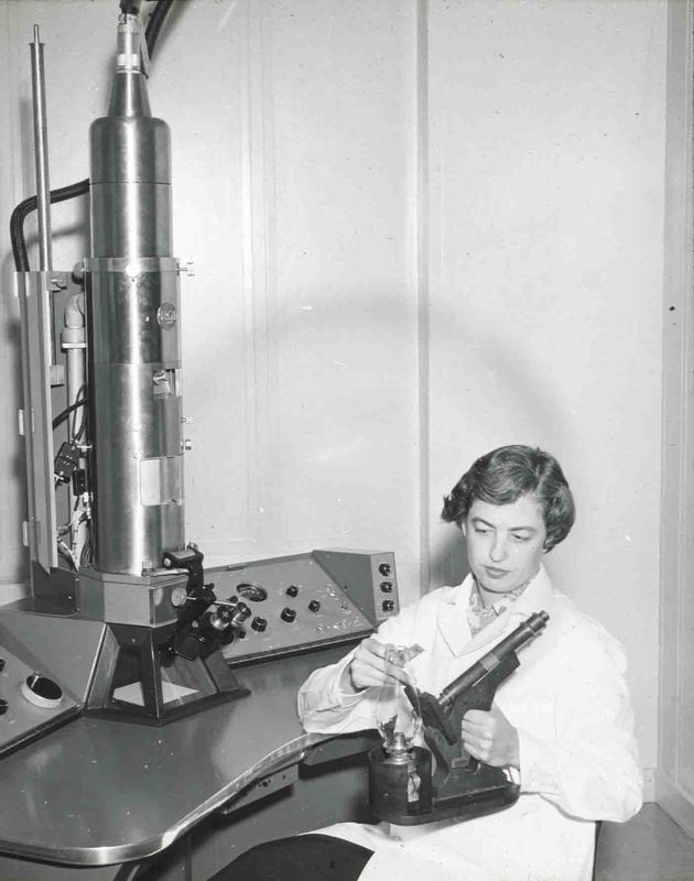
By 1960, the Department had four electron microscopes for the use of research staff, graduate students, postdoctoral fellows, and some medical students. Fawcett characterized the research endeavors of the Department as “mainly under the heading of cell biology, but they also include projects in such diverse fields as comparative anatomy, developmental biology, environmental physiology, endocrinology, neuroanatomy, reproductive physiology, and radiobiology.”
Don W. Fawcett recognized that gross anatomy continued in importance, particularly for teaching, but the amount of time devoted to its study in the curriculum had to decrease due to pressure from other subjects. He echoed Oliver Wendell Holmes of a century earlier, stating, “The observant anatomists of the past have left little of human anatomy undiscovered in the four hundred years since Vesalius, but recent progress of thoracic and orthopedic surgery has given point and clinical significance to structural relations that, in the past, were only of academic interest. Work is being done by one our clinical associates to meet the continuing need for reinvestigation of the applied anatomy of certain regions to support technical advances in surgery.”


