Anatomy
Oliver Wendell Holmes made and acquired preparations and casts of human and animal anatomy for use in his lectures. These are some of those casts and preparations that he donated to the Warren Anatomical Museum.
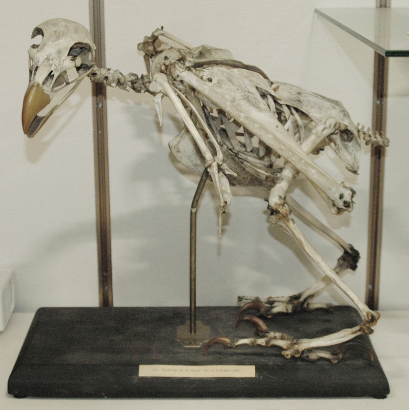
This eagle skeleton was mounted and prepared in the early-to-mid 19th century. It was donated by Oliver Wendell Holmes to the Warren Anatomical Museum in 1851.
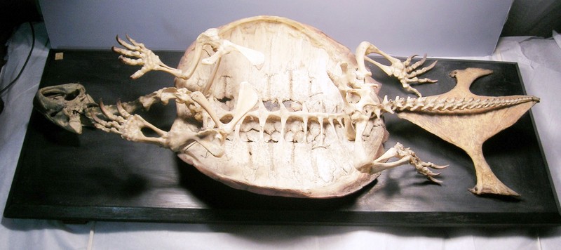
This snapping turtle skeleton was mounted and prepared in the early-to-mid 19th century. It was donated by Oliver Wendell Holmes to the Warren Anatomical Museum in 1855 along with the turtle's lungs, dried and inflated.
This anatomical specimen was dissected out, prepared, and mounted by Oliver Wendel Holmes between 1830 - 1849 to display the first two intercostal arteries. Holmes donated the preparation to the Warren Anatomical Museum in 1849.
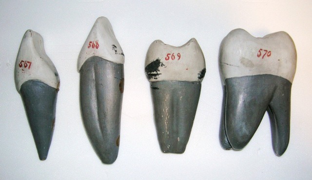
These tooth models were built in the early-to-mid 19th century and used by Oliver Wendell Holmes to teach dental anatomy. Holmes donated them to the Warren Anatomical Museum in 1851.
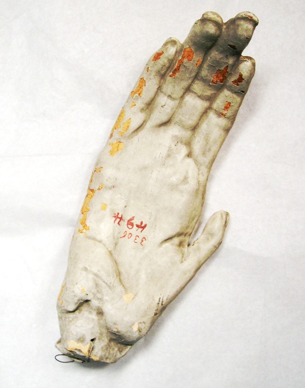
This chimpanzee hand was cast in plaster by Jefferies Wyman between 1830 and 1851. Wyman gave the cast to Holmes, who donated in to the Warren Anatomical Museum in 1851.
This sinus preparation was dissected out, injected and dried by Richard Hodges between 1845 and 1874. Hodges gave the preparation to Oliver Wendell Holmes, who donated it to the Warren Anatomical Museum in 1874.
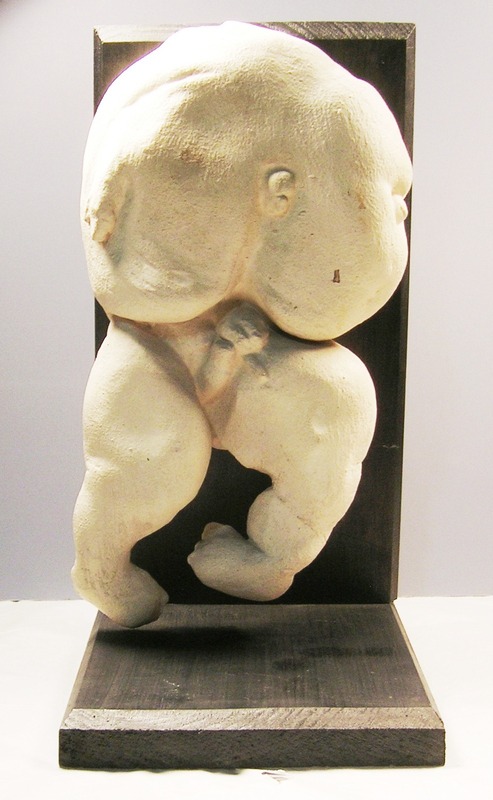
This plaster cast of an acephalic fetus was taken by Oliver Wendell Holmes in June 1837. The fetus and its healthy twin were born on June 25, 1837 to a patient of Ward N. Boylston. The subject did not survive. The cast, the subject's skeleton, its small intestine, a lithographic drawing of the subject and color drawing of the subject's circulatory system by Jefferies Wyman were donated to the Boston Society for Medical Improvement between 1837 - 1848. The cast was acquired by the Warren Anatomical Museum when the pathological museum of the Boston Society for Medical Improvement was transferred to Harvard Medical School circa 1871.

