Professor
Border lines of knowledge, in some provinces of medical science is a published and somewhat expanded version of Holmes’ introductory lecture to the students at Harvard Medical School at the opening of term on November 6, 1861. Although he refers to human anatomy as “an almost exhausted science,” Holmes goes on to list some of his own anatomical observations: “The nucleated cells found connected with the cancellated structure of the bones, which I first pointed out and had figured in 1847, and have shown yearly from that time to the present, and fossa masseterica, a shallow concavity on the ramus muscle, which acquires significance when examined by the side of the deep cavity on the corresponding part in some carnivore to which it answers, may perhaps be claimed as deserving attention. I have also pleased myself by making a special group of the six radiating muscles which diverge from the spine of the axis, or second cervical vertebra, and by giving to it the name stella musculosa nuchæ. But this scanty catalogue is only an evidence that one may teach long and see little that has not been noted by those who have gone before him.”
This dissected and dried preparation of the muscles of the cervical vertebrae named by Holmes stella musculosa nuchae (“muscular star of the neck”) is on display in the Warren Anatomical Museum.
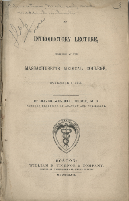
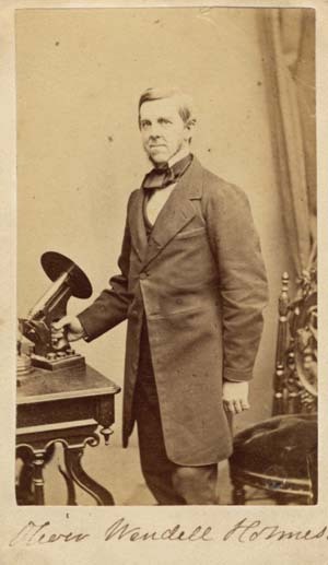
Holmes assumed the professorship of anatomy and physiology at Harvard in the fall of 1847 and delivered this introductory lecture to the students on November 3. In an overview of the medical developments of Boston, Holmes alludes here to “the strange magic of the enchanted goblet”—the first administration of ether anesthesia in operative surgery just a year before: “The knife is searching for disease, the pulleys are dragging back dislocated limbs, nature herself is working out the primal curse which doomed the tenderest of her creatures to the sharpest of her trials, but the fierce extremity of suffering has been steeped in the waters of forgetfulness, and the deepest furrow in the knotted brow of agony has been smoothed forever.”
The lecture prompted The Boston medical and surgical journal to aver that “it is the best discourse ever delivered in the Medical School of Harvard University. It abounds with bright thoughts, and there is a kind of elasticity and vigor running through its pages, that refreshes the reader.”
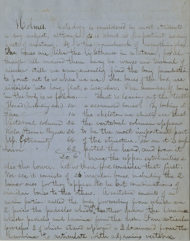
Edward Andem Whiston (1838-1909) of Framingham received a medical degree from Harvard in 1861, then served as a surgeon with the 1st and 16th regiments of the Massachusetts Volunteer Infantry during the Civil War. He was later the port physician of Boston and a founder and manager of the Newton Hospital. These are some of his notes on Holmes’ introductory lectures during the fall term at Harvard, probably from 1858.
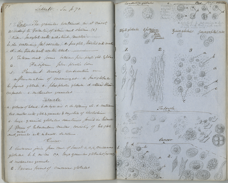
Holmes used these notes and outlines to deliver his course of lectures during the winter and spring terms. The notebook also includes a series of microscopic demonstrations made in April and May, 1851.
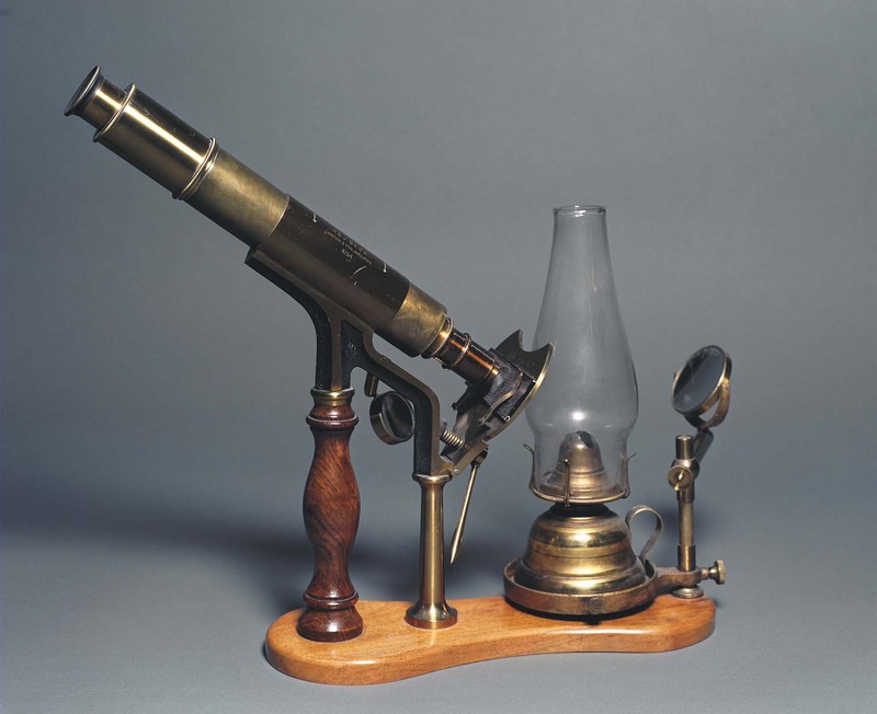
Holmes was offering practical instruction in the use of the microscope to medical students at Harvard by 1855. In an address to the Boston Microscopical Society in 1877, Holmes said, “My dealing with the instrument has been principally as a teacher, and not of microscopy as a specialty, but as a fractional portion of long-extended courses on anatomy, delivered to large classes. The most I could hope for was to teach them the rudiments of histology, and more especially to give them knowledge enough to make them wish for more. I have therefore aimed at having perfectly and easily manageable instruments, at selecting the more important and interesting objects, and at making everything as plain as practicable, knowing well that if a mistake in looking through a microscope is within the bounds of possibility, the young student will be certain to make it.”
A classroom demonstration brass monocular microscope with mirror and oil lamp, used by Oliver Wendell Holmes. It is mounted onto a wooden base with a heavy wooden handle.
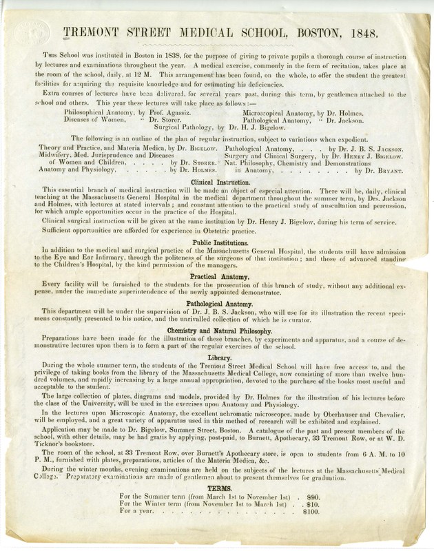
Holmes was one of the founders and faculty members of the Tremont Street Medical School; he offered courses in anatomy, physiology, and, as attested by this prospectus for the 1848 course, regular instruction in microscopic anatomy, and was one of the first physicians in America to do so. The microscopic demonstrations are described in an 1853 catalogue of the Tremont School: "These will be given by Dr. Holmes, weekly, through the Spring months…. They will illustrate by recent specimens, and a large number of carefully selected English and French preparations, the leading facts of animal and vegetable structure and development, and will be enforced by practical tests of the students' knowledge."


